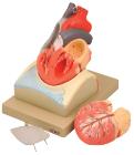
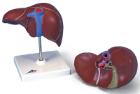
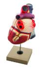
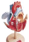
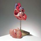
Enlarged, sectioned so that both ventricles and atria open to expose the valves. Large blood vessels near the heart and musculature of the heart are shown. Separates into 4 parts. On base.
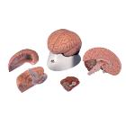
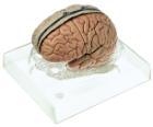
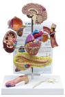
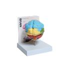
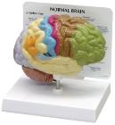
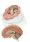
Model Brain 4-Part, right half can be disassembled into: Frontal with parietal lobes, Brain stem with temporal and occipital lobes, Half of cerebellum, great educational tool for the human nervous system and anatomy of the brain, Dimensions : 14 x 14 x 17.5 cm
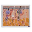
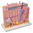
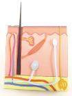
Skin 3 Part Model, The model consists of three individual parts on a common stand that represent sections of the human skin with a magnification of 80x
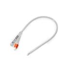
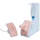
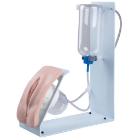
Simulator Catheterisation-Model, male, Ideal for demonstrating disposable & ballon catheters as well as suprapubic aspiration. Can be dismantled into two parts, natural size, made of special plastic. On a stand with green base, LxWXH: 18 X 18 X 30 cm
Sectioned through the ventricles and auricles. The bicuspid and tricuspid semilunar and sigmoid valves are shown. Separates into 3 parts. Mounted on base
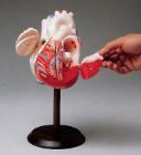
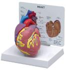
Model-Heart two parts, 100cm tall, Communicate the basics of heart anatomy to beginning biology students, Colorful and durable, features both external and internal components, including valves, two-piece model is mounted on a stand and can be removed for up-close study
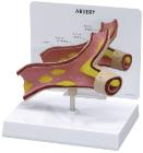
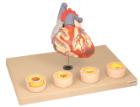
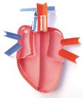
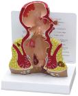
Heart dissected in two parts shows the thickened ventricle walls and cardiac valves. Four individual blood vessel model show coronary artery disease/ atherosclerosis and progression in the blood vessel, myocardial infarction and damage.
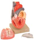
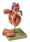
Life size model dissectible in 2 parts. The anterior heart wall can be removed to show the left and right ventricles and atria as well as the tricuspid, pulmonary, mitral and aortic valves. Mounted on base.
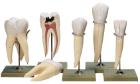
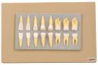
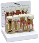
Enlarged approximately 2 times, set of 16 teeth cast in break resistant material having accurate anatomical details. Complete set as in half of upper & lower jaw
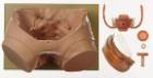
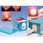
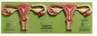
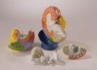
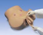
Model For Gynecological Patient Education, This unique gynecological training model is ideal for demonstration purposes and for realistic insertion of female barrier contraceptive devices, which are placed in the vaginal/cervical area.
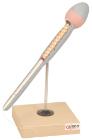
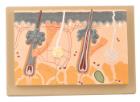
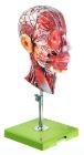
Enlarged 70 Times, full size, shows section through three layers of the hair covered skin of the head. Also shows hair follicles with sebaceous, sweat glands, receptors, nerves and vessels.
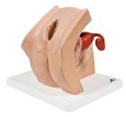
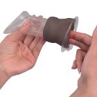
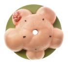
Model Female Condom Dark Skin, Dimensions: 12 cm, shows the labia and vagina up to the cervix in a simplified representation for didactic reasons, and is used for demonstrating and learning the insertion of a female condom
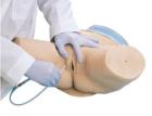
BASIC catheterization trainer set, both male and female bladder catheterization procedures can be realistically demonstrated, practiced and assessed. The easy-to-change genital inserted into the holder and held in place with magnets, with Both inserts
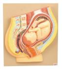
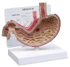
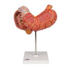
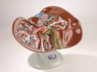
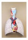
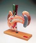
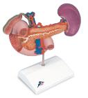
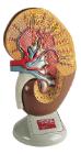
Rear Organs Of The Upper Abdomen, The upper abdomen organ model shows the duodenum (partially opened), gall bladder (opened) and bile ducts (opened), the pancreas (revealing large ducts), the spleen and the surrounding vessels in natural size